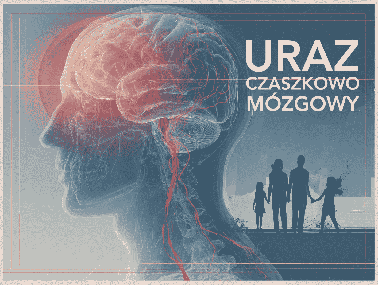
Traumatic brain injury (TBI) – rehabilitation – a comprehensive guide for families
NORMAN Neurological Rehabilitation Centre
Therapy results
What patients say about us
Legal notice: this content is for information only and does not constitute medical advice. If you have any health concerns, please consult a doctor.
The epidemiology of traumatic brain injury and the scale of the problem
Traumatic brain injury (TBI) is a global health problem and one of the leading causes of death and neurological disability. It is estimated that there are more than 69 million cases of TBI worldwide each year. In the United States, TBI accounts for hundreds of thousands of hospital admissions and tens of thousands of deaths every year. The true scale of the problem is in fact even greater, because the statistics do not capture all the milder injuries treated on an outpatient basis or those that go unreported.
TBI affects people of all ages, but those especially at risk are older adults, young children, teenagers, military personnel and people over the age of 65. The most common cause of injury remains falls, while in younger patients road traffic accidents, sports injuries and violence are also significant. Men suffer traumatic brain injuries more often and more often die as a result of them.
The significance of TBI extends far beyond the period of acute treatment, because a brain injury can result in long-term cognitive and memory impairments, emotional and behavioural changes, and neurological deficits. In many patients this affects the ability to live independently, return to work and maintain social relationships, and so it also places a burden on families and carers. Increasingly, TBI is seen not as a one-off event but as a disease process that can last a lifetime.
Research shows that people who have had a moderate or severe TBI have a reduced life expectancy and an increased risk of death from secondary causes, such as seizures, infections or neurological complications. More than half of survivors still struggle with moderate or severe disability, and a significant proportion need help from carers with everyday activities.
Classification and severity of TBI
The main tool for assessing the severity of an acute brain injury is the Glasgow Coma Scale (GCS), which rates the level of consciousness based on eye opening, verbal response and motor response. On this basis, TBI is classified into three main categories.
Mild TBI (GCS 13–15)
This group includes concussion, among others. Most often there are transient symptoms such as headache, nausea, confusion or retrograde amnesia. In some patients, post-concussion syndrome develops, with persistent headaches and difficulties with concentration or mood. Mild injuries make up the majority of all TBI cases.
Moderate TBI (GCS 9–12)
Moderate injuries involve a longer loss of consciousness or deeper confusion, along with neurological symptoms and abnormalities on imaging. They usually require hospital admission and observation, and sometimes neurosurgical intervention. Many patients regain their independence with the right therapy, but moderate deficits often remain.
Severe TBI (GCS 8 or below)
A severe injury means deep unconsciousness or coma and is often associated with a direct threat to life due to extensive haematomas, massive brain swelling or damage to the brainstem. It requires immediate intensive treatment, often surgery and a stay in intensive care. The outlook is serious, and mortality remains high.
The Glasgow Coma Scale does not, however, capture the full complexity of the injury. A complete assessment is also shaped by the results of a CT or MRI scan, the presence of haematomas, skull fractures and brain swelling, as well as the overall clinical course. In practice, therefore, a combination of the GCS, imaging and observation of the patient is used.
Modern medicine is moving towards a more multidimensional classification of TBI. Increasing emphasis is placed on the profile of clinical symptoms, imaging results, biomarkers of nerve tissue damage, and non-medical factors such as age, coexisting conditions and the context of the injury. The aim is to better predict the course of the illness and to select treatment more precisely.
The pathophysiology of primary and secondary injury
A traumatic brain injury sets off a complex cascade of pathological changes. A distinction is made between primary injury, which occurs directly at the moment the mechanical force is applied, and secondary injury, which develops in the minutes, hours and days after the event. Primary injuries include, among others, brain contusions, intracranial haematomas, diffuse axonal injury and penetrating wounds.
- Cerebral ischaemia and hypoxia: any episode of low oxygen or low blood pressure dramatically worsens the outlook, because the injured brain is especially sensitive to a lack of oxygen and energy substrates.
- Brain swelling and raised intracranial pressure: increasing swelling disrupts blood flow to the brain and can lead to dangerous shifts of brain structures.
- Excitotoxicity and oxidative stress: the excessive release of glutamate, the influx of calcium into neurons and the formation of free radicals accelerate the death of nerve cells.
- Inflammatory response: the activation of microglia and astrocytes and the influx of white blood cells support repair processes, but excessive inflammation can further damage brain tissue.
- Metabolic and systemic dysfunction: hormonal, fluid and electrolyte, glucose and sympathetic disturbances affect the outlook and the course of the acute phase of treatment.
The strategy for acute treatment focuses on breaking this chain of secondary injury: preventing hypoxia and low blood pressure, controlling swelling and raised intracranial pressure, protecting neurons from excitotoxicity and free radicals, and limiting an excessive inflammatory response.
Diagnosis and neurological assessment
The diagnosis of TBI relies on a parallel clinical assessment of the patient and imaging tests. The neurological examination assesses the level of consciousness, the pupils’ response to light, the strength and symmetry of limb movements, the reflexes and the mechanism of injury. As early as the pre-hospital stage, simplified consciousness scales are used and care is taken to secure the basic vital functions.
The gold standard of imaging in acute head injury remains a non-contrast CT scan of the brain. CT quickly detects haematomas, skull fractures, larger contusions, brain swelling and signs of raised intracranial pressure. In patients with at least a moderate TBI, the scan should be performed as soon as possible.
Magnetic resonance imaging (MRI) is more sensitive than CT in detecting certain changes, especially diffuse axonal injury, small haemorrhagic foci and brainstem injuries. It is usually performed once the patient has been stabilised, in the subacute or chronic phase, when the extent of the damage needs to be assessed more precisely.
In severe injuries, invasive monitoring of intracranial pressure (ICP) plays an important role, and sometimes so does ventricular drainage, which allows cerebrospinal fluid to be drained off. Specialist centres also use multimodal monitoring, such as CPP, PbtO2, continuous EEG or cerebral microdialysis.
Post-injury biomarkers such as GFAP and UCH-L1 are becoming increasingly important. Their raised levels in the blood may support the assessment of brain damage and, in the future, help to better select patients for further diagnosis and treatment.
Acute treatment in hospital and in intensive care
The pre-hospital phase and the emergency department
Right at the scene, the ABC principle applies: securing the airway, assessing breathing and supporting circulation. The aim is to prevent hypoxia and low blood pressure, which dramatically worsen the outlook. A patient with a suspected severe TBI should be taken as quickly as possible to a centre with neurosurgery and intensive care.
In the emergency department, stabilisation of vital functions, neurological assessment and imaging are carried out in parallel. If the CT scan reveals changes requiring surgical intervention, a decision is made to operate urgently.
- A large epidural or subdural haematoma with a mass effect and a shift of brain structures.
- An expanding focus of brain contusion or another lesion with a significant mass effect.
- Acute post-traumatic hydrocephalus, for example after intraventricular haemorrhage.
- High intracranial pressure that is resistant to conservative treatment.
Neurosurgical management includes, among other things, a craniotomy to evacuate haematomas, the placement of a ventricular drain and, in extreme situations, a decompressive craniectomy. Decompression surgery can be life-saving but does not always improve the final functional outcome, which is why these decisions are made on an individual basis.
Intensive care
- Blood pressure: low blood pressure must be avoided and systolic values kept in line with the Brain Trauma Foundation guidelines, in order to protect blood flow to the brain.
- Intracranial pressure (ICP): treatment for raised intracranial pressure is usually started when values exceed 22 mm Hg.
- Cerebral perfusion pressure (CPP): the aim is to maintain a range of around 60–70 mm Hg to ensure adequate blood flow through the brain.
- Oxygenation and ventilation: normal oxygen saturation and normocapnia are maintained, and hyperventilation is used only briefly as an emergency measure.
Treatment of brain swelling and raised intracranial pressure
- A body position with the head raised by around 30 degrees, to improve venous drainage from the brain.
- Sedation and analgesia, which reduce the brain’s metabolism and limit spikes in ICP caused by agitation, pain or coughing.
- Osmotic therapy, for example mannitol or hypertonic saline, to reduce brain swelling.
- Drainage of cerebrospinal fluid in patients who have a ventricular drain in place.
- Therapeutic hypothermia and barbiturates, considered in selected cases of resistant raised intracranial pressure.
- Decompressive craniectomy in situations where other methods fail.
General medical management in intensive care
- Early enteral feeding to limit catabolism and the risk of complications.
- Anticonvulsant prophylaxis during the first week after a severe TBI, usually using phenytoin or levetiracetam.
- Prevention of infection, aseptic care and early diagnosis of infection.
- Prevention of venous thromboembolism using mechanical methods and heparin, when the patient’s condition allows.
- Parallel treatment of any coexisting multi-organ injuries and systemic complications.
- Neuroprotective drug therapy has limited scope; one exception can be tranexamic acid used early in selected patients with haemorrhagic TBI, whereas steroids are contraindicated.
Acute treatment of TBI has three basic goals: to protect the brain from secondary injury, to stabilise the patient and to prepare them for further rehabilitation. The first days after the injury are critical, and the outcome of treatment depends both on the extent of the injury and on the quality of intensive medical care.
Rehabilitation after injury and post-hospital care
Rehabilitation begins as early as possible, often already in intensive care, where the first measures are put in place to prevent complications from immobility. Once stabilised, the patient is transferred to a neurological rehabilitation ward or a specialist post-injury programme.
- Physical therapy: improving muscle strength, range of motion and coordination, together with therapy for gait and balance disorders, as well as preventing contractures and muscle wasting.
- Speech and swallowing therapy: working on speech, comprehension, language expression and safe swallowing, including specialist therapy for speech and dysphagia.
- Neuropsychology and cognitive training: exercising memory, attention and executive functions, and educating the family about behavioural and communication difficulties.
- Psychological and psychiatric support: help with depression, anxiety, behavioural disturbances, emotional lability and readjustment to a new life situation.
- Orthopaedic aids and assistive technologies: orthoses, wheelchairs, standing equipment, robotic rehabilitation systems and other tools chosen to suit the patient’s needs.
The duration of rehabilitation varies greatly. The greatest progress is usually seen in the first months after the injury, but the brain’s neuroplasticity allows for improvement over the longer term as well. After discharge from hospital, rehabilitation should be continued on an outpatient basis or during residential neurological rehabilitation stays.
The family plays an enormous role in the recovery process. They should be involved in rehabilitation, learn how to support the patient’s independence and discover ways of communicating in the case of speech, memory or behavioural difficulties — much as with loss of speech after a stroke and aphasia. In many cases, education is also needed on care, nutrition, tracheostomy or enteral feeding.
Return to the community and readjustment
The ultimate stage of rehabilitation is the widest possible reintegration of the patient into society: a return to work, school, family life and social relationships. Some patients need vocational training, educational support, adaptation of their workplace or an individualised course of study. In severe cases, the priority is to maximise independence in the basic areas and to prepare the family for long-term care.
Sometimes it is necessary to adapt the home to the needs of a person with a disability, and in the most severe cases a stay in a nursing and care facility or long-term care at home is considered. Legal and social matters are also important, such as a disability certificate, a disability pension, respite care or funding for rehabilitation equipment.
Long-term consequences and chronic care
- Cognitive impairment: problems with memory, concentration, thinking speed, planning and organising the day.
- Lasting changes in personality and behaviour: irritability, impulsivity, apathy, withdrawal, difficulties in relationships and in controlling emotions.
- Post-traumatic epilepsy: the risk of seizures rises especially after severe injuries and can persist for years.
- Motor and sensory disorders: paresis, ataxia, visual disturbances and other deficits that make independent functioning harder, including paresis of the hand and upper limb.
- Hormonal complications: damage to the hypothalamus or pituitary gland can lead to endocrine disorders and require hormone replacement therapy.
- Mental health problems: depression, anxiety, PTSD, addiction and chronic difficulties with adjustment are common after TBI.
- Neurodegenerative risk: a severe TBI can increase the risk of dementia and other neurodegenerative conditions later in life.
Long-term care of a person after TBI is best provided by an interdisciplinary team: a family doctor, a neurologist, a rehabilitation physician, a physiotherapist, a psychologist and a psychiatrist and, where needed, also an endocrinologist, a speech and language therapist and a social worker. Only such an approach makes it possible to respond to the many-sided consequences of the injury.
The outlook in TBI depends on the extent of the primary injury, the quality of acute treatment and the intensity of rehabilitation. Many patients with a moderate injury return to relatively independent living, though often with some limitations. In severe injuries the outlook can be uncertain, but the support of the family and access to appropriate care are enormously important. It is precisely long-term treatment, rehabilitation and the informed involvement of loved ones that lay the foundation for the best possible quality of life after an injury.
References
- Dewan MC, Rattani A, Gupta S, et al. Estimating the global incidence of traumatic brain injury. Journal of Neurosurgery. 2018;130(4):1080–1097. DOI:10.3171/2017.10.JNS17352.
- Centers for Disease Control and Prevention (CDC), National Center for Injury Prevention and Control. Traumatic Brain Injury in the United States: Epidemiology and Rehabilitation. CDC Report to Congress, 2015. Atlanta, GA: Centers for Disease Control and Prevention.
- Carney N, Totten AM, O’Reilly C, et al. Guidelines for the Management of Severe Traumatic Brain Injury, Fourth Edition. Neurosurgery. 2017;80(1):6–15. DOI:10.1227/NEU.0000000000001432.
- CRASH-3 trial collaborators (Roberts I, et al.). Effects of tranexamic acid on death, disability, vascular occlusive events and other morbidities in patients with acute traumatic brain injury (CRASH-3): a randomised, placebo-controlled trial. The Lancet. 2019;394(10210):1713–1723. DOI:10.1016/S0140-6736(19)32233-0.
- Manley GT, Dams-O’Connor K, et al. A new characterization of acute traumatic brain injury: The NIH-NINDS TBI Classification and Nomenclature Initiative. Lancet Neurology. 2025;24(6):512–523. DOI:10.1016/S1474-4422(23)00164-9.
- Centers for Disease Control and Prevention (CDC). TBI Data – TBI-Related Hospitalizations and Deaths (web resource). National Center for Injury Prevention and Control, 2024. (accessed: 2025).
- Chou A, Torres-Espin A, Krukowski K, et al. Empowering data sharing and analytics through the Open Data Commons for Traumatic Brain Injury (ODC-TBI) research. Neurotrauma Reports. 2022;3(1):139–157. DOI:10.1089/neur.2021.0061.
- Masel BE, DeWitt DS. Traumatic brain injury: a disease process, not an event. Journal of Neurotrauma. 2010;27(8):1529–1540. DOI:10.1089/neu.2010.1358.
- Yue JK, Deng H. Traumatic Brain Injury: Contemporary Challenges and the Path to Progress. Journal of Clinical Medicine. 2023;12(9):3283. DOI:10.3390/jcm12093283.
Read next
Brain tumour – symptoms, treatment, surgery and rehabilitation (a complete guide)
Brain tumour – symptoms, diagnosis, surgery and rehabilitation step by step. Find out what a brain tumour means and what effective treatment looks like. A practical guide for patients and families.
Read more →
Multiple sclerosis (MS) – rehabilitation – a guide for patients and carers
Multiple sclerosis (MS) is a chronic, autoimmune neurological disease in which the immune system attacks the body’s own nerve cells, leading to damage of the myelin sheaths surrounding neurons. MS most often affects young adults between the ages of 20 and 40, and is twice as common in women as in men. The disease follows a highly varied course – most patients experience alternating periods of relapses (flare-ups) and remission,...
Read more →
Speech therapy after a stroke – a guide and speech exercises
A practical guide for the families of stroke patients: how to understand aphasia, how to exercise the facial muscles and speech, and how to talk to your loved one.
Read more →
Loss of speech after a stroke. Aphasia and the role of the speech therapist in the therapy process.
Aphasia after a stroke is the loss of the ability to understand and produce speech. We explain the types of aphasia, the role of the speech therapist and the directions rehabilitation can take.
Read more →
Can every stroke patient be rehabilitated?
Whether rehabilitation after a stroke is possible depends on the patient’s health, the time since the event and the right choice of therapy. We explain when rehabilitation is possible and how to find the right course of action.
Read more →
When should rehabilitation after a stroke begin?
Early rehabilitation after a stroke usually starts on the neurology ward, but its scope and pace always depend on the patient’s condition and the doctor’s decision.
Read more →
Stroke – brain areas and the effects of brain damage.
The brain is the source of our identity, intellect and emotions. Here is a short guide to the different areas of the brain and the deficits that can arise when they are damaged by an ischaemic stroke.
Read more →
Stroke – emotions, mood and depression
A stroke can damage the brain structures responsible for regulating emotions and mood, which is why depression, anxiety, motivation problems and difficulties with rehabilitation can appear afterwards.
Read more →
Stroke symptoms: how to recognise them and how to respond?
Recognising the symptoms of a stroke quickly and calling for help immediately can save a life and reduce the risk of lasting complications.
Read more →
The next step
Let's talk
We are here to answer any questions you may have about the rehabilitation process.
Describe your case
Send us the details of the patient's condition and we will assess them from a therapeutic perspective.
Email consultation
Write to us describing the condition and the patient's current state of health. We will reply with our view of your situation in the context of rehabilitation.
Video of the patient
You can send us a video showing the patient's current condition. We will respond to your situation and explain the therapeutic options available.